Macular Hole
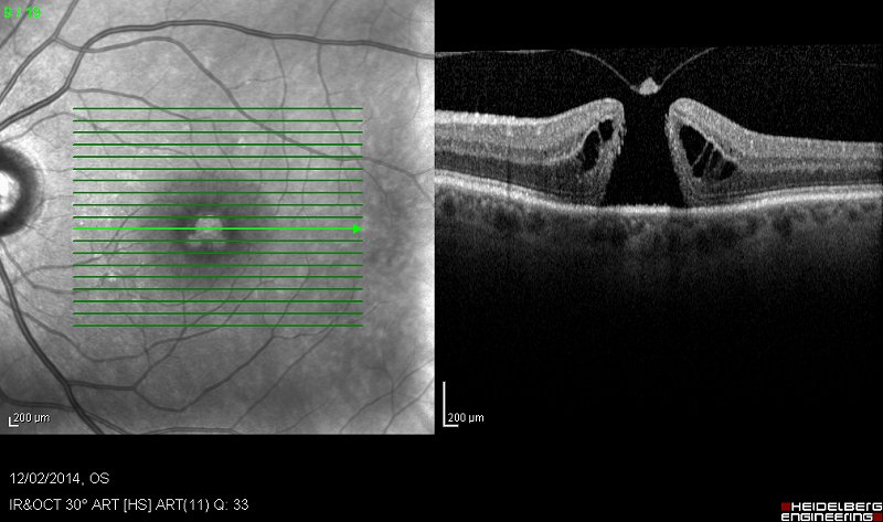
 MACULAR HOLES can occur in the highly sensitive area of the retina called the macula, which is used for fine vision and reading. Although the exact reason for this is not known, they are believed to be caused by the vitreous pulling on the macular.
What are the symptoms?
Central vision usually deteriorates and reading becomes difficult with some patients complaining of visual distortion particularly when looking at straight lines such as a pole or edge of a table.
How is a macular hole diagnosed?
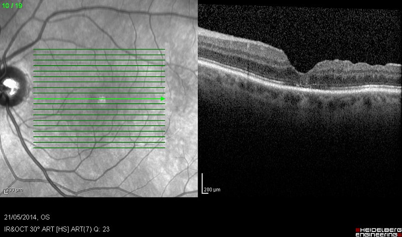
Inspection of the retina by a retinal expert, and a non-invasive scan called an OCT, will confirm the condition.
What is the treatment?
 This depends on the stage of the macular hole. While very early holes may be self-limiting, surgery is usually required in most patients who have significant symptoms. Macular hole surgery is carried out as a day case. Three needle-sized holes are made in the white of the eye allowing removal of the vitreous. The hole is repaired and the eye cavity is filled with a temporary gas bubble to seal the hole. As most eyes develop a cataract after surgery, it is common practice to carry out cataract surgery at the same time. The whole operation takes approximately 60-90 minutes and is carried out under local anaesthetic. Stitches are not required. At the end of the operation a pad is applied to protect the eye. Patients may be asked to keep their head in a face down position for a few days. Called 'posturing' it facilitates the gas bubble to seal the macular hole. Further details on post-operative care are explained during consultation and assessment.
Eye drops will reduce any inflammation and prevent infection. It is normal for the eye to feel watery, itching and experience mild discomfort for a while after macular hole or any retinal surgery. As the gas bubble dissolves gradually over the ensuing weeks, vision will steadily improve. In most cases your eye will take about six weeks to heal and then you will need to see your optician for glasses.
Small macular holes may be treated with Ocriplasmin injection in to the eye. This new treatment is less invasive and may be a suitable alternative to vitrectomy.


